


A C-arm machine is helpful for a variety of facilities. A C-arm machine is a cutting-edge medical tool that employs X-ray technology to scan various body sections and generates excellent images. A C-arm machine must be used accurately and cautiously. It cannot be very safe to consider all there is to know about C-arm machines. This gadget is an investment. Therefore you want to choose wisely. You can select the ideal C-arm machine for your facility if you have as much knowledge about them as possible. Their versatility makes them suitable for any facility. While doing precise tasks, C-arm devices provide clinicians with confidence. If you’re ready to invest in a C-arm, ensure the model will suit your needs now and in the future. Consider the following important factors: Talk with your medical physicist. They can help ensure the C-arm setup is safe and advise you on monitoring and radiation safety training issues.
Power Consumption
C-arms require a generator for powering the X-ray tube and image intensifier. Choosing a low-powered one can help you save up to 15% of the radiographer’s workload. It allows the surgical team to work faster while reducing patient and theatre staff risk. The C-arm dimensions and maneuverability are also important considerations. Look for units that are compact enough to fit into your room and allow you to rotate them around a patient without hitting anything else in the room. Depending on the procedures you perform, choose an Orthoscan FD-OR C-arm that offers high resolution for optimal imaging quality. You can also select a device with advanced functions like digital subtraction angiography, which removes bone and soft tissue to visualize blood vessels. This feature is particularly helpful for pain management physicians who need to access tiny nerve pathways.
Image Quality
When choosing a c-arm image, quality is one of the first things to consider. The c-arm unit is a large digital black-and-white camera that takes fluoro/x-ray anatomy images. The image intensifier converts the X-rays into high-intensity light to produce brighter images. It leads to better contrast, sharpness, and resolution. The pixel count is another important factor in determining c-arm image quality. The higher the pixel count, the more details an image can show.
Additionally, higher pixel counts can help reduce skin entry dose. Additionally, c-arms feature reference features such as color-coded axes to make repositioning faster and easier. It increases precision and safety. It also helps prevent misalignment during a procedure. For even better image quality, the tube’s height can be adjusted.
Patient Comfort
Whether setting pedicle screws or a stent graft, surgeons depend on C-arms to help them visualize intricate structures during spine procedures. They also depend on surgical tables designed specifically for use with c-arms. A sense of patient comfort is a dynamic state that can include relief from pain and emotional distress and an emerging sense of strength, confidence, value, safety and control in their interaction with healthcare providers. Efforts to promote comfort include empathetic listening, reassuring touch and careful use of humor/chit-chat. To get the most from a C-arm, call in the expertise of a medical physicist to assess your facility’s radiation safety levels and monitor personnel with radiation badges.
Convenience
For those primarily involved in orthopedic, urology, and vascular procedures, a C-arm with a 9″ image intensifier will be sufficient. When selecting a new C-arm, evaluating its functionality and convenience will help you to choose the right one for your needs. For example, the latest units allow wireless image transfer to your facility’s PACS system. This feature reduces wear and tear on the equipment. It saves time by eliminating the need to physically take a heavy piece of equipment out of the procedure room and down a hallway to upload surgical images (though getting a mini c-arm versus a larger one still makes wheeling these between rooms when necessary easy). Another important feature is the ability to perform vascular and neurology imaging. It will allow you to use your C-arm for more surgical procedures than you could before. Further, convenience is found in the orbital rotation capability of your C-arm. If the detector area can rotate, be elevated, or put closer to the ground to match a patient’s needs, this can save a lot of uncomfortable repositioning for the client, especially if the machine can come to them.
Warranty
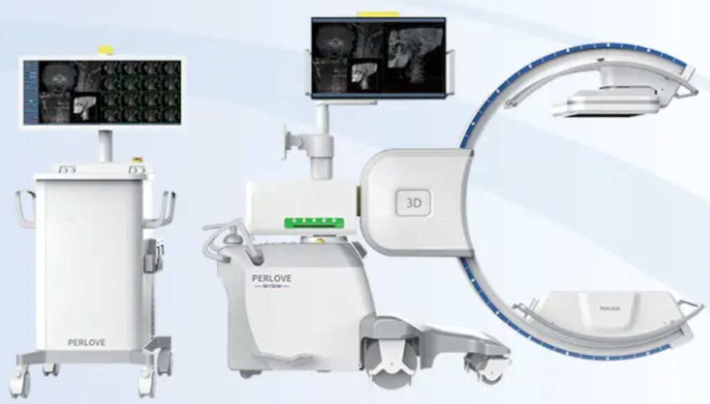
Whether or not you decide to buy your C-arm through an OEM, it’s important to look at the warranty. For example, a good warranty should cover both parts (including glassware) and labor and offer a full plan rather than a prorated one. Assessing your facility’s current procedures and determining what image quality you need, such as 2D or cone-beam CT (3D). It’s crucial to comprehend the primary components of a C-arm machine. The X-ray generator and source are the two direct parts. A C-shaped arch links these two primary portions together. There is a workstation where the operator can operate the apparatus in addition to the X-ray source and X-ray generator. Look at the device’s versatility to do whatever your practice needs. Facilities should consider purchasing a model with an isocentric design, which saves time by reducing the need to move patients and equipment around. Finally, look into trade-in programs to ensure the device can be upgraded.